

Optometrist and Butterfly Conservation member Simon Berry explains his remarkable research into how nocturnal insects’ eyes adapt
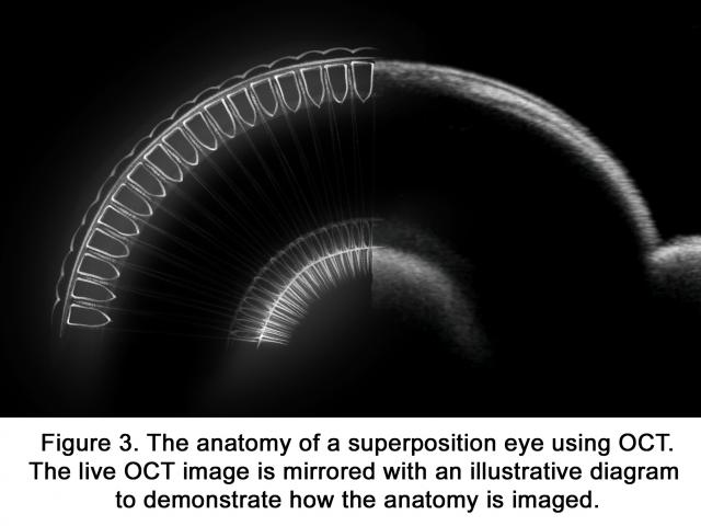
I’ve always been interested in photography and capturing things that are unusual or have never been seen before. At my optometry practice, we use a specialist bit of equipment called an OCT (Ocular Coherence Tomography) scanner, which shows a cross section of biological tissue. This 3D scanner, sometimes likened to optical ultrasound, is very useful in diagnosing certain eye conditions.
A few years ago, I became interested in whether this scanner could be used to scan insect eyes, and wondered if it could reveal something new about them. In 2018 I started scanning butterfly eyes – I managed to scan 12 different species of our local butterflies found in Durham. This is the first time anyone has ever scanned a compound eye with an OCT scanner. For someone used to looking at these scans, the scans do look unusual, but they didn’t really show anything new about butterfly anatomy. Then in 2019, I started scanning moth’s eyes, and they turned out to be more interesting.
Adaptation
To see effectively at night, moths and other nocturnal insects have a problem they must overcome. When light levels are low, their eyes need to be very sensitive; but they also need a way of adapting to environmental light conditions, and protecting those sensitive organs, if they encounter a bright light.
Human eyes have a pupil that changes size to regulate the amount of light entering the eye. Moths have a different method – their eyes have a light-absorbing pigment that changes position to limit the light getting in. This pigment migration is difficult to record because it is a dynamic process and only occurs in a live moth.
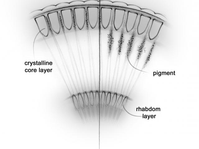
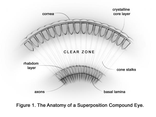
When a moth is ‘dark adapted’, the pigment is squeezed in between the crystalline cone layer of the eye. This allows the maximum possible light to pass through the eye to the retina. When the insect becomes ‘light adapted’, the pigment migrates into a clear zone within the eye, blocking the light and reducing the amount of light reaching the moth retina.
We know about this process of light adaption in moths because they have a tapetum (similar to a cat’s eye) which means that moths’ eyes glow when light is shone directly at them. The state of adaption can be measured by how brightly their eyes glow.
The only other way of seeing the pigment migration in a moth is to look at the eye of a dead insect under a microscope.
New ground
By using OCT technology on insect eyes for the first time, my research has shown that this can be used to visualise the structures and processes within the compound eye of a live moth. The big advantage of this is that the results can be viewed in real time, and the process does not harm the insect.
This should hopefully lead to a better understanding. For example, the process of light and dark adaption in a moth is relatively slow. This means that a light adapted moth flying around a light source is disadvantaged if it moves away from that light source, because they will not be able to resolve the same detail until they become dark adapted again. They are effectively caught by that light source.
Using this technology will hopefully help us to explain other aspects of moth behaviour.
Find out more
Simon had an article on this research published in Environmental Entomology (Volume 51, Issue 4, August 2022).
Butterfly Conservation scientists are also at the forefront of understanding the impact of LED lights on months.